
VIP member
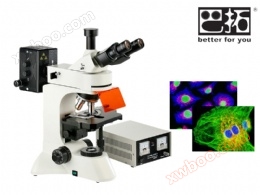
BSF-58 infinite fluorescence microscope mercury lamp model
BSF-58 infinite fluorescence microscope mercury lamp model
Product details
BSF-58 fluorescence microscope is suitable for fluorescence microscopy and transmission bright field observation The optical system adopts an infinitely far flat field achromatic fluorescent objective lens without magnification chromatic aberration and a large field eyepiece, with clear and bright imaging and a wide field of view. The body design that meets ergonomic requirements makes it more comfortable and easy for you to operate. It is an ideal instrument for research in biology, cytology, oncology, genetics, immunology and other fields, and can be used by scientific research, universities, epidemic prevention and other departments.
Product Features:
▲ Large field of view eyepiece, with a field of view diameter of 22mm and a large and flat field of view.
▲ Fluorescence falling observation, modular design, easy replacement of fluorescence modules.
▲ The infinite high beam path system adopts an infinite far flat field achromatic objective lens, which has excellent imaging effect.
▲ Ergonomic design, unique dial style fine adjustment (right-hand).
▲ Translucent and reflective lighting can observe opaque, semi transparent, and transparent objects.
▲ The V-type adopts a 6.3 megapixel SONY high-sensitivity chip camera, suitable for fluorescence photography.
System Introduction:

Technical specifications:
|
Technical Specifications |
||
|
eyepiece |
Large field of view WF10X (field of view size Φ 22mm) |
|
|
Infinite flat field achromatic fluorescent objective lens |
PL 4X/0.10 working distance: 19.8 mm |
|
|
PL 10X/0.25 working distance: 5.0 mm |
||
|
PL FL40X/0.85 (spring) working distance: 0.42 mm (no magnification color difference) |
||
|
PL 100X/1.25 (spring, oil) working distance: 0.36mm |
||
|
Eyepiece tube |
Triple eyepiece, tilted at 30 degrees |
|
|
Falling fluorescent lighting system |
Power box 110V/230V selectable |
|
|
Mercury lamp 100W/DC |
||
|
Rotating fluorescent falling light illuminating device (including ultraviolet, ultraviolet, blue, and green excitation filter groups) |
Ultraviolet (UV) excitation wavelength: 320-380nm, emission wavelength: 435nm |
|
|
Purple (V) excitation wavelength: 380-415nm, emission wavelength: 475nm |
||
|
Blue (B) excitation wavelength: 450-490, emission wavelength: 515nm |
||
|
Green (G) excitation wavelength: 495-555, emission wavelength: 595nm |
||
|
focusing mechanism |
Coarse micro coaxial focusing, micro grid value: 2 μ m, with locking and limiting devices |
|
|
converter |
Five holes (inward ball positioning) |
|
|
stage |
Double layer mechanical mobile type (size: 210mmX140mm, range of movement: 75mmX50mm) |
|
|
Transmissive lighting system |
Abbe spotlight NA.1.25 can be raised and lowered up and down |
|
|
Blue filter and frosted glass |
||
|
Collector, suitable for halogen lighting (with built-in field of view light barrier) |
||
|
6V 30W halogen lamp, adjustable brightness |
||
noteThe standard imaging system of the system is a 6.3 million SONY chip high-sensitivity camera. Due to the special nature of fluorescence photography, if you need a cooled CCD camera, please contact customer service
Imaging system:6.3 million high-sensitivity microscope camera (USB 3.0)
This series of cameras uses Sony Exmor, Exmor R, Exmor RS back illuminated CMOS sensors. The EXmor series CMOS sensors adopt double-layer noise reduction technology, which has ultra-high sensitivity and ultra-low noise.
thisThe camera integrates a 12 bit ultra fine hardware image signal processor and video streaming engine((Ultra-fineTMHISPVP)Implementing hardware through HISPVPDemosaic, Adjustment, automatic exposure, gain adjustment, one click white balance, image color adjustment, saturation adjustment, gamma correction, brightness adjustment, contrast adjustment, Bayer format image to RAW data for final 8/12Bit output. HISPVP has transferred the traditional CPU processing to hardware processing, greatly improving the camera's transmission speedReduced CPU usage.
Utilizing USB 3.0 data transfer technology to achieve fast and stable video transmission.
thisThe camera randomly provides advanced video and image processing application software ImageView; Provide Windows/Linux/OSX multi platform SDK; Supports native C/C++, C#/VB. Net, Directshow, Twain API。
thisThe above characteristics of the series camera can be used for capturing microscopic images in dark field, ordinary bright field, weak light or fluorescence light field.
thisThe basic characteristics of the camera series are as follows:
l adoptSony ExmorC-interface CMOS for CMOS back illuminated sensorsUSB3.0Camera;
l Adopting parallel A/D conversion technology to achieve ultra-low noise and low power consumption;
l The hardware resolution spans 3.1M~20M and other types;
l Zinc aluminum alloy precisionCNCshell
l USB 3.0 interface ensures high transmission speed;
l Ultra-FineTMThe hardware IPS video streaming engine ensures accurate color reproduction and provides advanced video and image processing application software quickly with the camera;
l Provide Windows/Linux/OSX multi platform SDK, supporting native C/C++, C#/VB. Net, Directshow, Twain API;
Imaging system parameters:
|
Product Model |
6.3 million USB 3.0 high-sensitivity microscope camera system |
|
sensor |
SONY 6.3M/IMX178(C) |
|
Chip size |
1/1.8“(7.37x4.92) |
|
Image Processor |
Ultra-FineTMHardware IPS video streaming engine |
|
像素大小 (um) |
2.4x2.4 |
|
Frame rate @ resolution |
38@1536x 1024 |
|
30@3072 x2048 |
|
|
exposure time |
0.1ms~15s |
|
Exposure control |
manual |
|
white balance |
ROI white balance/manual Temp Tit adjustment |
|
Color temperature control |
manual |
|
Settings |
White balance, exposure time, noise reduction, gain, gamma, etc |
|
Operating temperature |
-10~ 50℃ |
|
Operating humidity |
30~80RH |
|
operating system |
Microsoft® Windows®XP/ Vista / 7 / 8 /10(32 & 64 位) |
|
Optical interface |
Standard C interface |
|
data interface |
USB3.0 |
|
Capture/Control API |
Native C/C++, C #/VB.net, Directshow, Twain, and Labview |
|
Recording method |
Images and videos |
|
Cooling method* |
natural cooling |
|
power supply |
USB socket power supply (shared data interface) |
BSF-58 fluorescence microscope is suitable for fluorescence microscopy and transmission bright field observation The optical system adopts an infinitely far flat field achromatic fluorescent objective lens without magnification chromatic aberration and a large field eyepiece, with clear and bright imaging and a wide field of view. The body design that meets ergonomic requirements makes it more comfortable and easy for you to operate. It is an ideal instrument for research in biology, cytology, oncology, genetics, immunology and other fields, and can be used by scientific research, universities, epidemic prevention and other departments.
Product Features:
▲ Large field of view eyepiece, with a field of view diameter of 22mm and a large and flat field of view.
▲ Fluorescence falling observation, modular design, easy replacement of fluorescence modules.
▲ The infinite high beam path system adopts an infinite far flat field achromatic objective lens, which has excellent imaging effect.
▲ Ergonomic design, unique dial style fine adjustment (right-hand).
▲ Translucent and reflective lighting can observe opaque, semi transparent, and transparent objects.
▲ The V-type adopts a 6.3 megapixel SONY high-sensitivity chip camera, suitable for fluorescence photography.
System Introduction:
Technical specifications:
|
Technical Specifications |
||
|
eyepiece |
Large field of view WF10X (field of view size Φ 22mm) |
|
|
Infinite flat field achromatic fluorescent objective lens |
PL 4X/0.10 working distance: 19.8 mm |
|
|
PL 10X/0.25 working distance: 5.0 mm |
||
|
PL FL40X/0.85 (spring) working distance: 0.42 mm (no magnification color difference) |
||
|
PL 100X/1.25 (spring, oil) working distance: 0.36mm |
||
|
Eyepiece tube |
Triple eyepiece, tilted at 30 degrees |
|
|
Falling fluorescent lighting system |
Power box 110V/230V selectable |
|
|
Mercury lamp 100W/DC |
||
|
Rotating fluorescent falling light illuminating device (including ultraviolet, ultraviolet, blue, and green excitation filter groups) |
Ultraviolet (UV) excitation wavelength: 320-380nm, emission wavelength: 435nm |
|
|
Purple (V) excitation wavelength: 380-415nm, emission wavelength: 475nm |
||
|
Blue (B) excitation wavelength: 450-490, emission wavelength: 515nm |
||
|
Green (G) excitation wavelength: 495-555, emission wavelength: 595nm |
||
|
focusing mechanism |
Coarse micro coaxial focusing, micro grid value: 2 μ m, with locking and limiting devices |
|
|
converter |
Five holes (inward ball positioning) |
|
|
stage |
Double layer mechanical mobile type (size: 210mmX140mm, range of movement: 75mmX50mm) |
|
|
Transmissive lighting system |
Abbe spotlight NA.1.25 can be raised and lowered up and down |
|
|
Blue filter and frosted glass |
||
|
Collector, suitable for halogen lighting (with built-in field of view light barrier) |
||
|
6V 30W halogen lamp, adjustable brightness |
||
noteThe standard imaging system of the system is a 6.3 million SONY chip high-sensitivity camera. Due to the special nature of fluorescence photography, if you need a cooled CCD camera, please contact customer service
Imaging system:6.3 million high-sensitivity microscope camera (USB 3.0)
This series of cameras uses Sony Exmor, Exmor R, Exmor RS back illuminated CMOS sensors. The EXmor series CMOS sensors adopt double-layer noise reduction technology, which has ultra-high sensitivity and ultra-low noise.
thisThe camera integrates a 12 bit ultra fine hardware image signal processor and video streaming engine((Ultra-fineTMHISPVP)Implementing hardware through HISPVPDemosaic, Adjustment, automatic exposure, gain adjustment, one click white balance, image color adjustment, saturation adjustment, gamma correction, brightness adjustment, contrast adjustment, Bayer format image to RAW data for final 8/12Bit output. HISPVP has transferred the traditional CPU processing to hardware processing, greatly improving the camera's transmission speedReduced CPU usage.
Utilizing USB 3.0 data transfer technology to achieve fast and stable video transmission.
thisThe camera randomly provides advanced video and image processing application software ImageView; Provide Windows/Linux/OSX multi platform SDK; Supports native C/C++, C#/VB. Net, Directshow, Twain API。
thisThe above characteristics of the series camera can be used for capturing microscopic images in dark field, ordinary bright field, weak light or fluorescence light field.
thisThe basic characteristics of the camera series are as follows:
l adoptSony ExmorC-interface CMOS for CMOS back illuminated sensorsUSB3.0Camera;
l Adopting parallel A/D conversion technology to achieve ultra-low noise and low power consumption;
l The hardware resolution spans 3.1M~20M and other types;
l Zinc aluminum alloy precisionCNCshell
l USB 3.0 interface ensures high transmission speed;
l Ultra-FineTMThe hardware IPS video streaming engine ensures accurate color reproduction and provides advanced video and image processing application software quickly with the camera;
l Provide Windows/Linux/OSX multi platform SDK, supporting native C/C++, C#/VB. Net, Directshow, Twain API;
Imaging system parameters:
|
Product Model |
6.3 million USB 3.0 high-sensitivity microscope camera system |
|
sensor |
SONY 6.3M/IMX178(C) |
|
Chip size |
1/1.8“(7.37x4.92) |
|
Image Processor |
Ultra-FineTMHardware IPS video streaming engine |
|
像素大小 (um) |
2.4x2.4 |
|
Frame rate @ resolution |
38@1536x 1024 |
|
30@3072 x2048 |
|
|
exposure time |
0.1ms~15s |
|
Exposure control |
manual |
|
white balance |
ROI white balance/manual Temp Tit adjustment |
|
Color temperature control |
manual |
|
Settings |
White balance, exposure time, noise reduction, gain, gamma, etc |
|
Operating temperature |
-10~ 50℃ |
|
Operating humidity |
30~80RH |
|
operating system |
Microsoft® Windows®XP/ Vista / 7 / 8 /10(32 & 64 位) |
|
Optical interface |
Standard C interface |
|
data interface |
USB3.0 |
|
Capture/Control API |
Native C/C++, C #/VB.net, Directshow, Twain, and Labview |
|
Recording method |
Images and videos |
|
Cooling method* |
natural cooling |
|
power supply |
USB socket power supply (shared data interface) |
Online inquiry
-
Contacts
-
Company
-
Telephone
-
Email
-
WeChat
-
Verification Code
-
Message Content
-