
VIP member
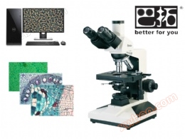
XSP-10C Trinocular Biological Microscope
XSP-10C Trinocular Biological Microscope
Product details
Instrument usage:
Batou®XSP-10CThe three eye biological microscope adopts a large field eyepiece and a flat field achromatic objective lens, which provides clearer imaging effects. This device not only allows visual observation, but also transmits the observed images to the computer screen through the camera, effectively relieving the fatigue of personnel who work for a long time. It can also conveniently save the observed images as pictures or videos, making it convenient for scientific researchers to record and archive samples
Batou®XSP-10CWidely used in microbiology, cytology, bacteriology, histology, medicinal chemistry and other research institutes, it can also be used as a medical test (non clinical), such as mite detection, skin sectioning, blood analysis, etc. It can be used for various biological experiments in the laboratory and for teaching purposes in schools.
Performance characteristics:
▲ Configure large field eyepiece and flat field achromatic objective lens,Clear imaging.
▲ 6V 20WHalogen lamp lighting,The brightness is adjustable.
▲ The three lens barrel allows for easy switching between eyepiece observation and camera observation,100%Spectral analysis, suitable for low light photography.
▲ Optional camera system, (500Choose freely between a 10 megapixel high pixel imager or a DSLR digital camera system.
Technical Specifications:
|
model |
XSP-10C |
|
brand |
|
|
eyepiece |
Broad VisionWF10X(Φ18mm)WF16X(Φ11mm) |
|
objective lens |
Flat field chromatic aberration correction4X/0.10 |
|
Flat field chromatic aberration correction10X/0.25 |
|
|
Flat field chromatic aberration correction40X/0.65(spring) |
|
|
Flat field chromatic aberration correction100X/1.25(spring,oil) |
|
|
magnification |
Visual inspection:40X,64X,100X,160X,400X,640X,*1000X,*1600X computer:100X-2600X(Configuration)500Ten megapixel camera with screen resolution640×480In the environment) |
|
Three eye tube |
binoculars(tilt30˚Adjustable pupillary distance),100%Split beam independent camera system. |
|
focusing mechanism |
Coarse micro coaxial focusing,Micro grid value:2Mm,Rough movement with adjustable elasticity,Equipped with locking and limiting devices,Add black plastic hand pillow board on the base |
|
converter |
four holes(Internal positioning of inward ball) |
|
stage |
Double layer mechanical mobile type(size:160mmX140mm,range of movement: 75mmX50mm) |
|
Abbe spotlight |
N.A.1.25Adjustable up and down |
|
Color filter |
Blue filter |
|
frosted glass |
|
|
Concentrator |
Suitable for halogen lamps |
|
light source |
6V 20W,halogen lamp,The brightness is adjustable. Customizable low heat white lightLEDLighting. |
Computer type/Composition of digital system:

Optional parts:
|
eyepiece |
Graduated eyepiece(0.1mm) |
|
|
converter |
five-hole(Internal positioning of inward ball) |
|
|
Color filter |
Green filter |
|
|
Yellow filter |
||
|
light source |
LEDConcentrator |
High brightness white light |
|
condenser |
dark field |
dry-type |
|
wet type |
||
|
Phase contrast device |
eyepiece |
centering telescope |
|
objective lens |
Phase contrast and color correction10X/0.25 PHP |
|
|
Phase contrast and color correction20X/0.40 PHP(spring) |
||
|
Phase contrast and color correction40X/0.65 PHP(spring) |
||
|
Phase contrast and color correction100X/1.25 PHP(spring,oil) |
||
|
Rotary phase contrast spotlight(PH-Ⅰ) |
||
|
Rotary phase contrast spotlight(PH-Ⅱ) |
||
|
Plug-in phase contrast spotlight |
||
|
Pull plate phase contrast spotlight |
||
VType imaging system parameters:
|
Product Model |
500ten thousandUSB2.0Microscopic camera system |
|
sensor |
Aptina CMOS 5.1M |
|
Chip size |
1/2.5“ (5.70x4.28) |
|
Image Processor |
Ultra-FineTMColor Processing Engine |
|
Pixel size(um) |
2.2x2.2 |
|
frame rate@resolution |
5@2592x1944 |
|
18@1280x960 |
|
|
60@640x480 |
|
|
scanning mode |
line-by-line scanning |
|
exposure time |
0.294ms~2000ms |
|
Exposure control |
manual/automatic |
|
white balance |
ROIwhite balance/manualTemp-Tintadjust |
|
Color temperature control |
manual/automatic |
|
Settings |
White balance, exposure time, noise reduction, gain, gamma, etc |
|
Operating temperature |
-10~ 50℃ |
|
Operating humidity |
30~80%RH |
|
operating system |
Microsoft® Windows®XP/ Vista / 7 / 8 /10(32 & 64bit) |
|
Optical interface |
standardCinterface |
|
data interface |
USB2.0 |
|
capture/controlAPI |
Native C/C++, C#/VB.NET, Directshow, TwainandLabview |
|
Recording method |
Images and videos |
|
Cooling method* |
natural cooling |
|
power supply |
USBPlug in power supply (shared data interface) |
Instrument usage: Batou®XSP-10CThe three eye biological microscope adopts a large field eyepiece and a flat field achromatic objective lens, which provides clearer imaging effects. This device not only allows visual observation, but also transmits the observed images to the computer screen through the camera, effectively relieving the fatigue of personnel who work for a long time. It can also conveniently save the observed images as pictures or videos, making it convenient for scientific researchers to record and archive samples Batou®XSP-10CWidely used in microbiology, cytology, bacteriology, histology, medicinal chemistry and other research institutes, it can also be used as a medical test (non clinical), such as mite detection, skin sectioning, blood analysis, etc. It can be used for various biological experiments in the laboratory and for teaching purposes in schools. Performance characteristics: ▲ Configure large field eyepiece and flat field achromatic objective lens,Clear imaging. ▲ 6V 20WHalogen lamp lighting,The brightness is adjustable. ▲ The three lens barrel allows for easy switching between eyepiece observation and camera observation,100%Spectral analysis, suitable for low light photography. ▲ Optional camera system, (500Choose freely between a 10 megapixel high pixel imager or a DSLR digital camera system.
Technical Specifications:
model XSP-10C brand eyepiece Broad VisionWF10X(Φ18mm)WF16X(Φ11mm) objective lens Flat field chromatic aberration correction4X/0.10 Flat field chromatic aberration correction10X/0.25 Flat field chromatic aberration correction40X/0.65(spring) Flat field chromatic aberration correction100X/1.25(spring,oil) magnification Visual inspection:40X,64X,100X,160X,400X,640X,*1000X,*1600X computer:100X-2600X(Configuration)500Ten megapixel camera with screen resolution640×480In the environment) Three eye tube binoculars(tilt30˚Adjustable pupillary distance),100%Split beam independent camera system. focusing mechanism Coarse micro coaxial focusing,Micro grid value:2Mm,Rough movement with adjustable elasticity,Equipped with locking and limiting devices,Add black plastic hand pillow board on the base converter four holes(Internal positioning of inward ball) stage Double layer mechanical mobile type(size:160mmX140mm,range of movement: 75mmX50mm) Abbe spotlight N.A.1.25Adjustable up and down Color filter Blue filter frosted glass Concentrator Suitable for halogen lamps light source 6V 20W,halogen lamp,The brightness is adjustable. Customizable low heat white lightLEDLighting.

Computer type/Composition of digital system:
Optional parts:
|
eyepiece |
Graduated eyepiece(0.1mm) |
|
|
converter |
five-hole(Internal positioning of inward ball) |
|
|
Color filter |
Green filter |
|
|
Yellow filter |
||
|
light source |
LEDConcentrator |
High brightness white light |
|
condenser |
dark field |
dry-type |
|
wet type |
||
|
Phase contrast device |
eyepiece |
centering telescope |
|
objective lens |
Phase contrast and color correction10X/0.25 PHP |
|
|
Phase contrast and color correction20X/0.40 PHP(spring) |
||
|
Phase contrast and color correction40X/0.65 PHP(spring) |
||
|
Phase contrast and color correction100X/1.25 PHP(spring,oil) |
||
|
Rotary phase contrast spotlight(PH-Ⅰ) |
||
|
Rotary phase contrast spotlight(PH-Ⅱ) |
||
|
Plug-in phase contrast spotlight |
||
|
Pull plate phase contrast spotlight |
||
VType imaging system parameters:
|
Product Model |
500ten thousandUSB2.0Microscopic camera system |
|
sensor |
Aptina CMOS 5.1M |
|
Chip size |
1/2.5“ (5.70x4.28) |
|
Image Processor |
Ultra-FineTMColor Processing Engine |
|
Pixel size(um) |
2.2x2.2 |
|
frame rate@resolution |
5@2592x1944 |
|
18@1280x960 |
|
|
60@640x480 |
|
|
scanning mode |
line-by-line scanning |
|
exposure time |
0.294ms~2000ms |
|
Exposure control |
manual/automatic |
|
white balance |
ROIwhite balance/manualTemp-Tintadjust |
|
Color temperature control |
manual/automatic |
|
Settings |
White balance, exposure time, noise reduction, gain, gamma, etc |
|
Operating temperature |
-10~ 50℃ |
|
Operating humidity |
30~80%RH |
|
operating system |
Microsoft® Windows®XP/ Vista / 7 / 8 /10(32 & 64bit) |
|
Optical interface |
standardCinterface |
|
data interface |
USB2.0 |
|
capture/controlAPI |
Native C/C++, C#/VB.NET, Directshow, TwainandLabview |
|
Recording method |
Images and videos |
|
Cooling method* |
natural cooling |
|
power supply |
USBPlug in power supply (shared data interface) |


Online inquiry
-
Contacts
-
Company
-
Telephone
-
Email
-
WeChat
-
Verification Code
-
Message Content
-